Page 32 - Nutrinsight-4
P. 32
NutrInsight • Satiety: News Insights
Neuroimaging results revealed that these signals are supported by different neural substrates: goal values are correlated with activity in the medial orbitofrontal cortex, decision values are correlated with activity in the central orbitofrontal cortex, and prediction errors are correlated with activity in the ventral striatum. An important application of these results is to neuropathologies involving decision-making deficits such as addiction. Knowing the precise value computations carried out in different regions of the brain could aid in the development of behavioural and/or pharmacotherapeutic interventions targeted at specific stages of diseases.
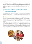
5.3 Bottom-up control of cognitive and emotional functions by nutrients
Figure 1 shows how the brain receives input from the internal milieu of the organism as well as from various cues from the environment, including food-associated cues. Studies of brain activation using fMRI have illustrated how signals coming from the internal milieu, and specifically from ingested nutrients, control cognitive and emotional functions. This “bottom-up” mechanism is illustrated in the following experiments.
Encoding food reward value
Associative learning allows an animal to predict future events, such as food reward, on the basis of sensory cues. One experimental paradigm to investigate associative learning is classical conditioning, as illustrated by the pioneer works of Pavlov. In classical conditioning, a previously neutral cue (the conditioned stimulus, or CS+) acquires biological significance after being paired with a natural reinforcer (the unconditioned stimulus, UCS). The progress of fMRI allows the neural substrate of such associations to be examined.
An fMRI study was carried out in 13 hungry human subjects during learning and anticipation of two food-based olfactory rewards (UCS) [Gottfried et al., 2003]. Arbitrary visual images were used as target and non-target CS+ stimuli, which were either paired (CS+p) or unpaired (CS+u) with the corresponding UCS. Another image was never paired with odors (CS-). During initial training, participants learned the picture-door associations while performing a visuospatial discrimination task. Significant neural responses in the posterior amygdala, rostromedial orbitofrontal cortex, ventral midbrain, primary olfactory cortex, insula, and hypothalamus indicated the contribution of these structures to the learning of picture-door contingencies (Figure 2).
32