Page 35 - Nutrinsight-4
P. 35
NutrInsight • Satiety: News Insights
Although the decision to eat and how much to eat can be considered a “free decision” in humans, unconscious processing appears to play an important role in shaping this decision. Using fRMI and statistical pattern recognition techniques, it has been shown that neural activity in the medial and lateral frontopolar cortex as well as the precuneus reflecting a future motor response (such as eating) precedes the time of the decision entering consciousness by as much as 10 seconds [Soon et al., 2008]. Since brain activity consistently preceded the conscious decision, it has been argued that the brain had already unconsciously made a decision to eat even before the subject became aware of it.
5.5 Weight loss and the “hungry brain”
The above examples show how neural activity in brain regions affecting regulatory and hedonic aspects of energy homeostasis can be investigated using fMRI. One interesting problem is that of the increased hunger and food intake that typically occur following attempts to lose weight in obese patients. It has been hypothesized that weight loss might induce changes in neural responsiveness making the “hungry brain” incapable of adequate control of food intake.
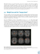
This was investigated in a group of obese patients [Rosenbaum et al., 2008]. Their neural activity elicited by food-related visual cues was examined before and after the loss of 10% of initial body weight. Following weight loss, predictable changes were observed in neural activity in brain areas known to be involved in the regulatory, emotional, and cognitive control of food intake. Figure 4 shows exaggerated activity induced by visual food stimuli after weight loss in the brainstem, globus pallidus, middle frontal gyrus, middle temporal gyrus, parahippocampal gyrus, and culmen. Some of these exaggerated brain responses were normalized after administration of leptin, suggesting that they were due to the low circulating leptin levels induced by the weight loss. Thus, the weight-reduced state can be viewed as a state of relative leptin-deficiency, while the obese state is characterized by leptin resistance.
Figure 4: Exaggerated activity of neural brain structures following a 10% weight loss in obese subjects.
Many of these exaggerated responses were normalized after administration of leptin. BS: brainstem; Cul: culmen; GF: fusiform gyrus; GP: globus pallidus; GTm: middle temporal gyrus; GHp: parahippocampal gyrus
Source: Rosenbaum et al., 2008
35